Researchers at the Massachusetts Institute of Technology have developed a new imaging technology that allows you to see the distribution of electrical signals in the nerve tissues of the brain. This technology promises to be invaluable in researching the processes of thought formation, sensation, and elucidating the causes of such brain diseases as Alzheimer’s disease, epilepsy, and others.
Modern MRI (magnetic resonance imaging) technologies already provide us with a wealth of information, but they can only provide a rough visualization of the areas of the brain activated by a particular external stimulus. To get a picture of activity at the level of individual neurons, which “communicate” with each other and work in groups to form thoughts and feelings, requires an instrument with a greater resolution than an MRI scanner.
Lack of a tool capable of capturing all 86 billion neurons of the brain with its “look” compels neurobiologists to study the nervous system of the simplest living creatures, various worms and larvae, whose number of neurons in the brain amounts to hundreds. In addition, such studies use very complex and slow technologies, such as the insertion of electrodes into the nerve tissues of the brain, through which electrical signals are read.
A new technology developed by the team of Professor Ed Boyden (Ed Boyden) is able to provide a more complete picture of brain activity than other existing methods. This technology uses a specially selected fluorescent protein that binds to the cell membrane of neurons and responds with its glow to electrical signals. It is the glow of this protein and allows you to accurately track the path of signals in neural circuits.
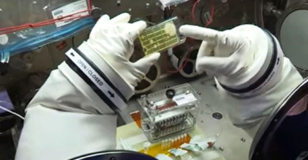
Professor Boyden’s group even had to build a specialized laboratory robot that selected the most suitable fluorescent protein from more than 10 million candidates. The robot absolutely independently injected each of the tested proteins into a tissue cell, grew the cells in petri dishes and took pictures of the results. Specialized software was used to analyze the results, which determined the location, luminosity and resistance of each protein to various adverse factors.
Finally, the robot managed to find the most suitable type of fluorescent protein, with which the researchers stained the nervous tissue of the worm Caenorhabditis elegans and brain tissue of the experimental rodent, which allowed them to get a visual picture of brain activity. This technology can be used in tandem with other optogenetic technologies that can either inhibit activity or, conversely, stimulate individual neurons, allowing them to determine the pathway of signals from these individual neurons.
With the new imaging technology at their disposal, the researchers are going to use it to make a detailed “activity map” of the entire brain of the experimental rodent. The data from this map will allow them in the future to identify with high precision the neural circuits and nodes responsible for processing certain stimuli, forming certain reactions and linking together areas of the brain that perform different functions.